Bioimaging core facility
About us
The UPOL-Bioimaging Facility (BF), formerly known as the UPOL-Microscopy Imaging Facility, is a critical pillar of the Institute of Molecular and Translational Medicine (IMTM), situated within the Medical Faculty of the University of Palacky, Olomouc. The facility primarily offers service-based access to sophisticated microscopy and other imaging technologies while promoting independent research initiatives conducted by its staff.
Our mission centers around providing cost-effective, shared access to these advanced technologies for a diverse user base. This includes the IMTM, Medical Faculty, University of Palacky, as well as external researchers from across the Czech Republic and beyond. We also uphold a key role in educating pre- and post-graduate students via numerous training programs and courses.
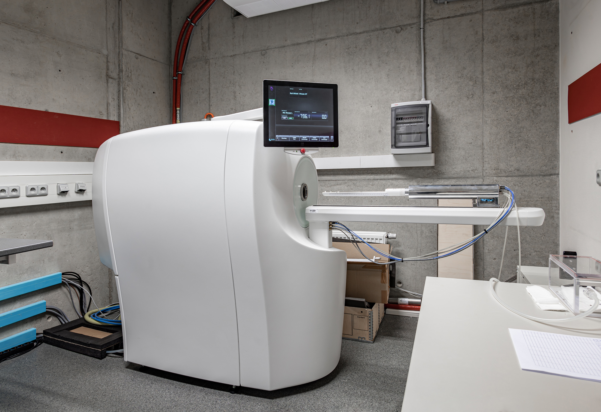
In Spring 2022, we broadened our capabilities to include a new unit specializing in PET/MRI imaging for small animals, notably mice, thereby strengthening our capacity in translational biomedical research.
The facility is segmented into four distinct units: i) Fluorescence, Confocal, and Super-resolution Microscopy, ii) Atomic Force Microscopy, iii) Raman and Infrared Microscopy, and iv) PET/MRI Imaging. Each unit is headed by a lead who is accountable to both the Director of IMTM, Dr. Marian Hajduch, and the Project Manager of the Czech-Bioimaging project at IMTM, Dr. Martin Mistrik.
Supported by Czech-BioImaging
National research infrastructure comprising 16 leading imaging facilities across 10 research institutions in Prague, Vestec, Brno, České Budějovice, and Olomouc. Czech-BioImaging uniquely combines open access, educational courses, funding opportunities, and close cooperation with industry.
Through three national Euro-BioImaging nodes, Czech Bioimaging is part of Euro-BioImaging ERIC and is included in the Roadmap of Large Research Infrastructures of the Czech Republic for the years 2023—2026.
⇒ Czech-BioImaging Access policy info
⇒ Open access form
⇒ E-learning test
Why to choose us:
- State-of-the-Art Equipment & Diverse Services: The UPOL-Bioimaging Facility houses a vast array of cutting-edge imaging technologies, from superresolution microscopy techniques like SIM and PALM to the recently added NanoScan PET/MRI 3T system. This breadth of offerings ensures that researchers can access a comprehensive range of imaging solutions serving to diverse scientific inquiries.
- Dedicated & Expert Staff: With a team of seasoned professionals spanning researchers, imaging specialists, and IT experts, users can expect world-class expertise and guidance. Each staff member is not only proficient in operating advanced technologies but also offers valuable insights for optimizing experiments.
- Strong Educational Commitment: Affiliated with the University of Palacky's Institute of Molecular and Translational Medicine, the facility plays a pivotal role in grooming the next generation of researchers. Through its hands-on training programs, it provides both pre-and post-graduate students with invaluable experiences using top-tier bioimaging technologies.
- Enhancing International Competitiveness: The facility's commitment to state-of-the-art technology and high-caliber research significantly elevates the Czech Republic's global prominence in bioimaging. By continually pushing the boundaries of what's possible, it helps position the nation at the forefront of biomedical research.
- Strategic Regional & Global Partnerships: Located in Olomouc, the UPOL-Bioimaging Facility not only bolsters the region's scientific infrastructure but also forms strategic alliances both nationally and internationally.
Superresolution Microscopy Techniques
Proficient in advanced microscopy techniques including Structured Illumination Microscopy (SIM) and Photoactivated Localization Microscopy (PALM).
Long-Term Live-Cell Imaging
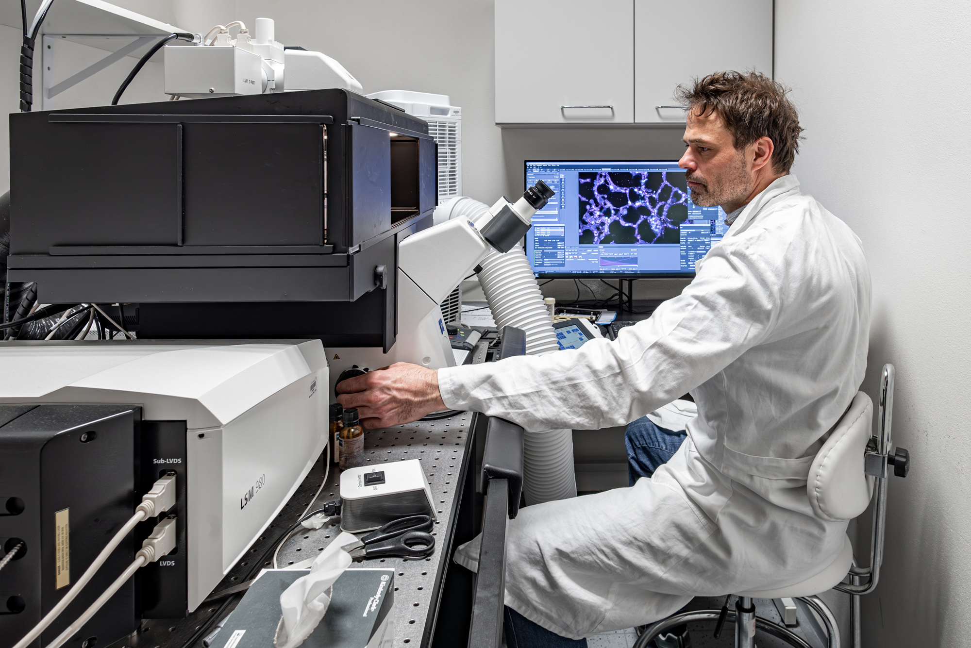
Utilizing dedicated spinning disk and laser scanning confocal microscopes for extended live-cell studies.
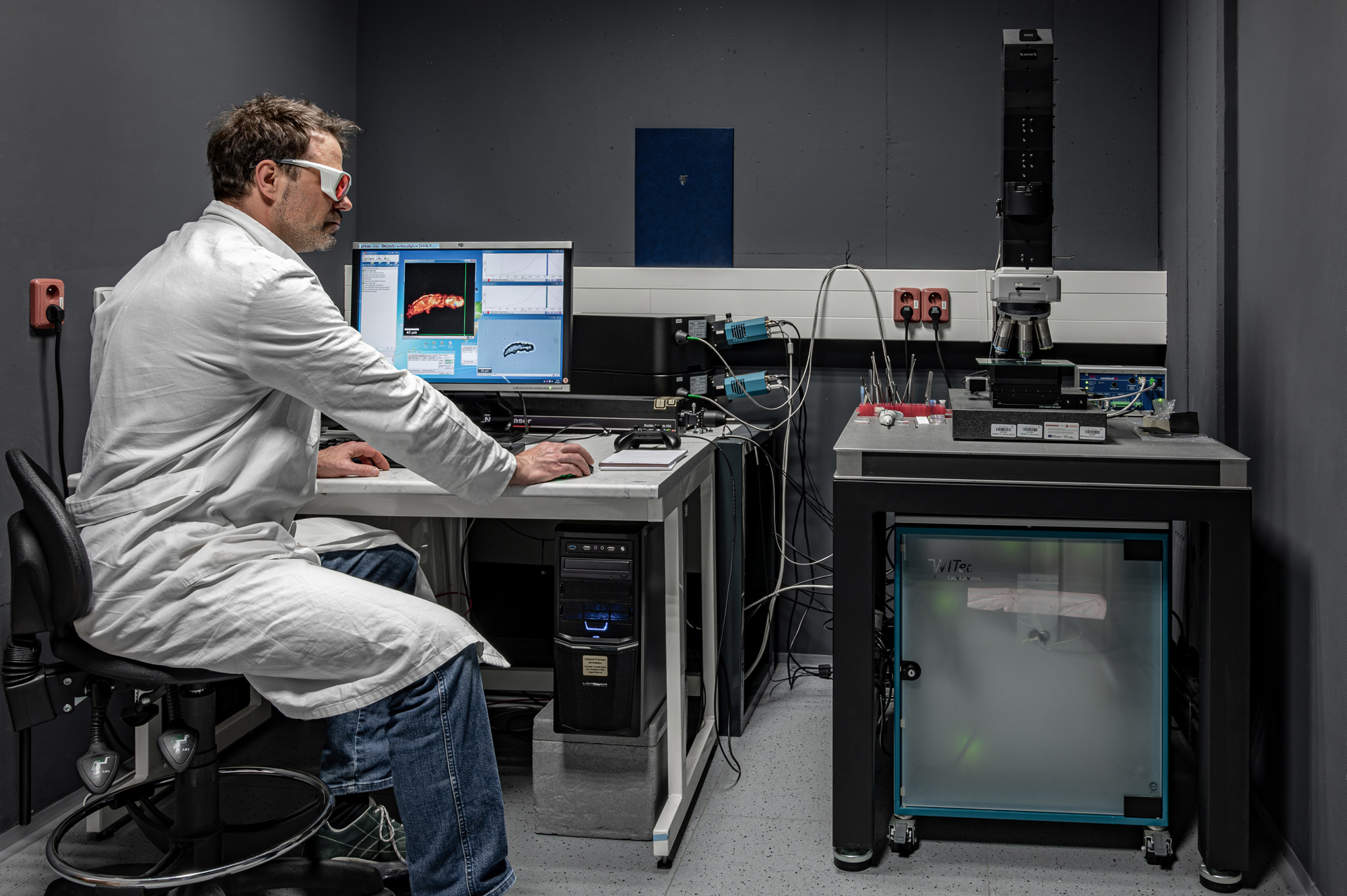
Specialized Laser Manipulation Techniques
Offers unique capabilities for inducing DNA damage in live cells. Additionally, the facility provides techniques for creating localized thermal damage at both cellular and subcellular levels.
Microscopic Environment Modifications
Custom-made modifications available for microscopic incubators to allow ambient temperature manipulation within sample chambers, ranging from 15-50°C.
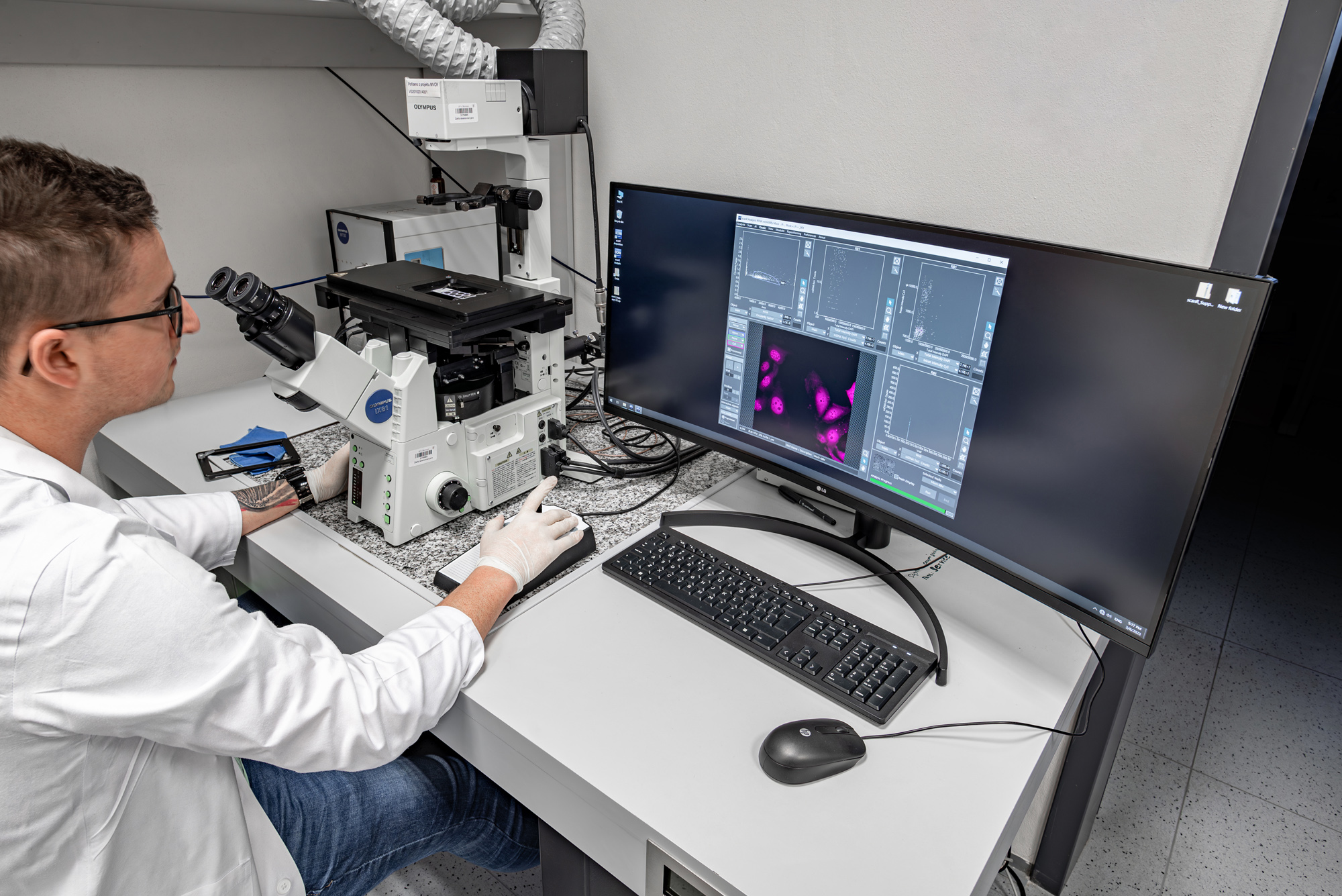
Quantitative Microscopy
Equipped with automated Olympus ScanR microscope designed for FLOW-cytometry-like analysis, and offering custom software solutions for comprehensive image analysis.
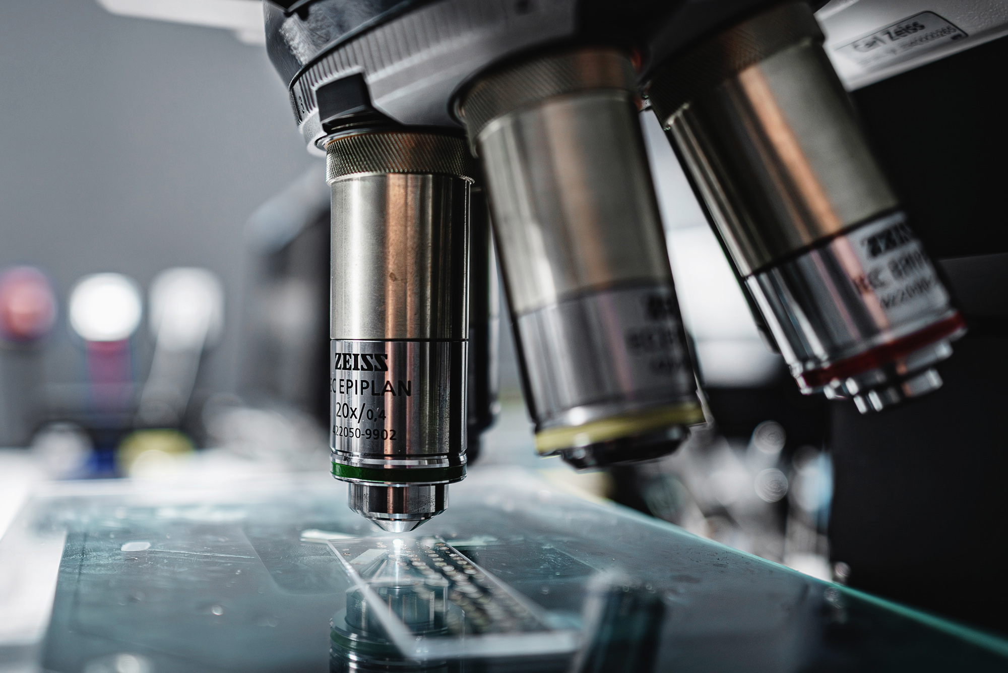
Microscopy-Based Analytical Methods
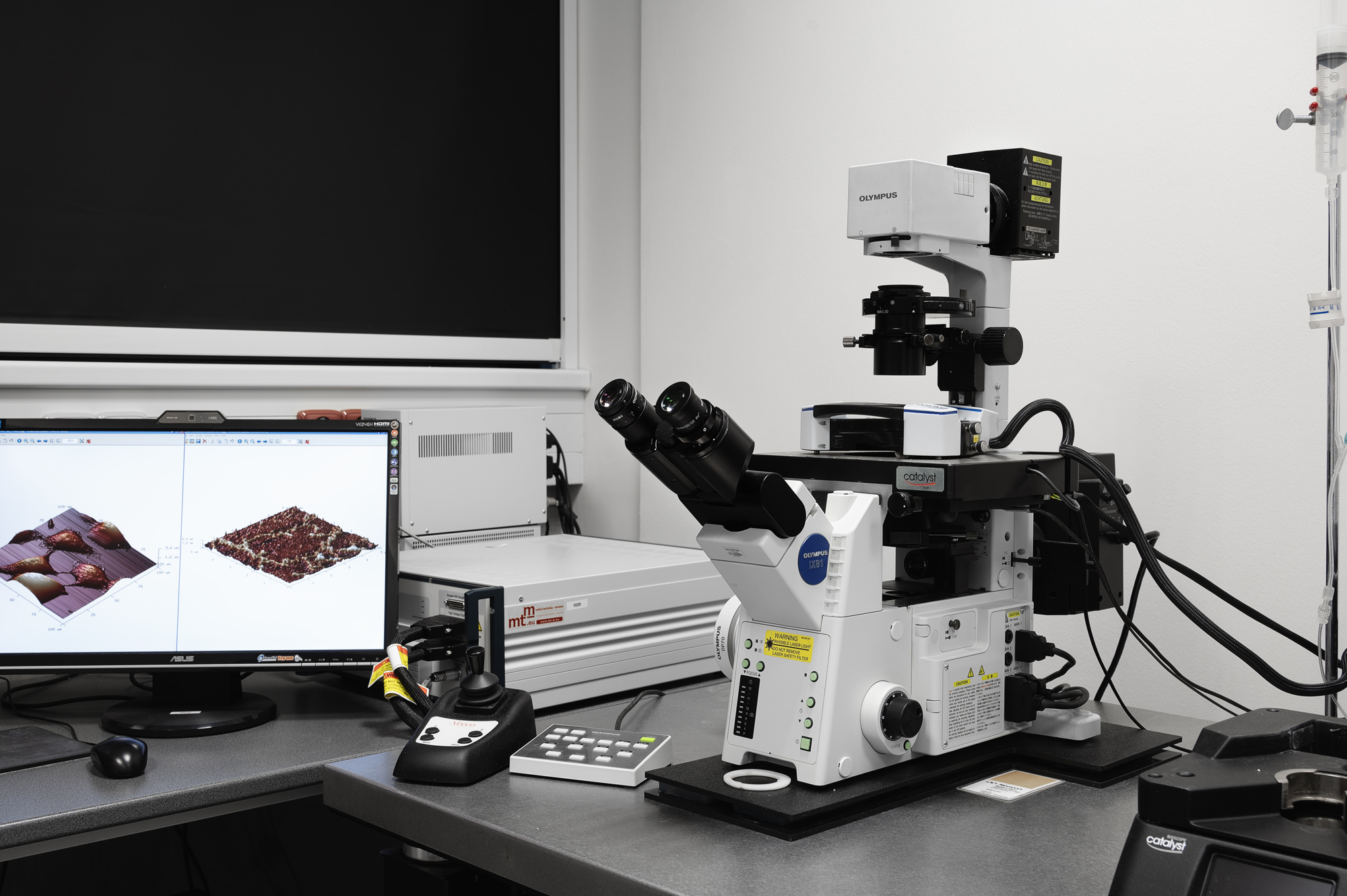
Wide range of analytical methods available for biocompatible materials, biological, and non-biological samples using technologies like Atomic Force Microscopy and stain-free microscopic techniques such as Raman and Infrared (IR) Microscopy.
Advanced PET/MRI Technology
A recently added service, the facility offers the NanoScan PET/MRI 3T system, crucial for precise imaging in small laboratory animals, primarily focusing on in vivo studies in mice and rats.
Spinning disk microscope
Olympus ScanR microscope
Zeiss microscope (LSM780, Elyra PS.1)
Zeiss LSM980
Olympus LSM microscope
NanoScan PET/MRI 3T Mediso
Confocal microscope Carl Zeiss LSM 780 with UV laser and ELYRA.PS1 module
Reserve this instrumentWITec alpha300 R + Confocal Raman imaging microscope system
Reserve this instrumentIf you have any questions or would like to discuss your genomics research needs, please don't hesitate to reach out to us. Our friendly and knowledgeable team is ready to assist you. You can contact us through the following methods:
- Phone: +420 585634873
- Email: martin.mistrik@upol.cz
- Location: Institute of Molecular and Translational Medicine, Faculty of Medicine and Dentistry and Czech Advanced Technology and Research Institute, Palacky University, Hnevotinska 5, 779 00 Olomouc, Czech Republic
We are excited to be a part of your genomics research journey and look forward to collaborating with you on groundbreaking discoveries that will shape the future of science and medicine. Let's unlock the potential of genomics together!
Head of the facility
Members
Photo gallery