Imaging and Tracing
The Imaging and Tracing group focuses on the preclinical development of novel radiopharmaceuticals and molecular imaging agents for the comprehensive evaluation of pathological conditions and monitoring of disease progression and therapy in vivo. This includes the development of cutting-edge radiolabelling and radioanalytical strategies, and the evaluation of novel candidate tracers in vitro and in vivo using a wide range of imaging approaches. We also develop small animal models of various diseases, particularly infections and cancer.
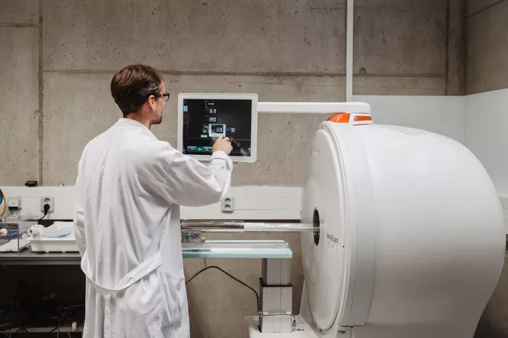
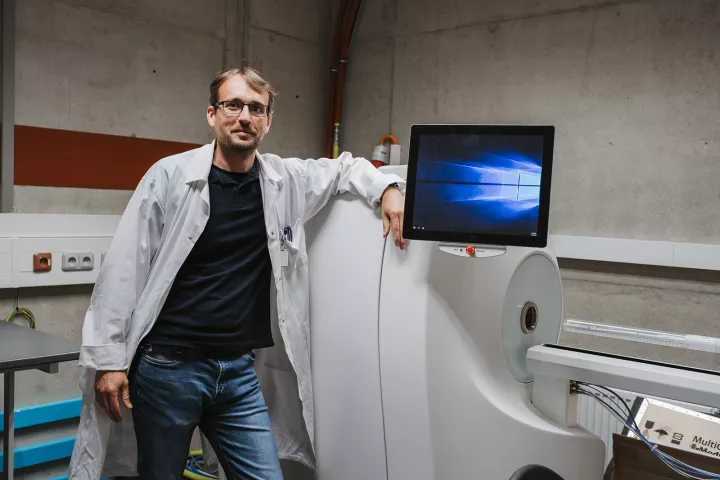
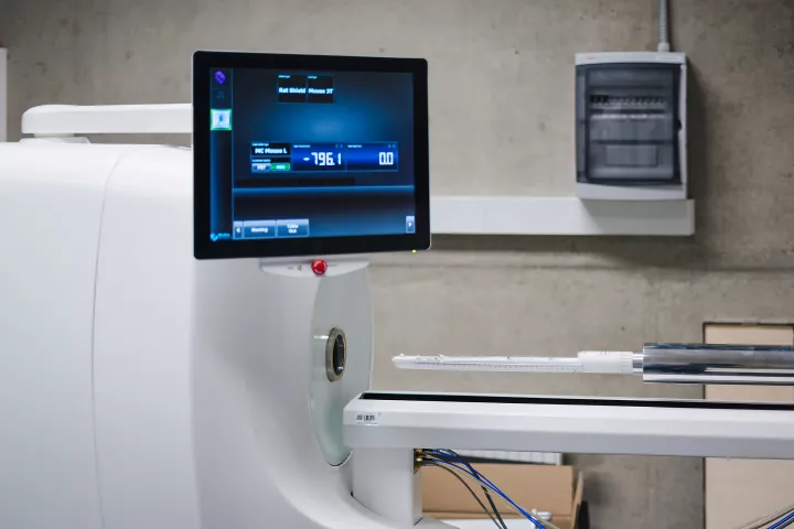
We perform non-invasive multimodal imaging and image analysis for the observation of various disease and biological models in living systems. Our state-of-the-art instrumentation currently includes an extensive suite of imaging modalities, such as positron emission tomography (PET), single photon emission computerized tomography (SPECT), computerized tomography (CT), magnetic resonance imaging (MRI), optical imaging (including bioluminescence and fluorescence ranging from the green to near-infrared region) and ultrasound (US). In addition, fully equipped radiochemistry laboratory and dedicated housing for animals undergoing longitudinal imaging studies is available.
- Preclinical development of novel radiopharmaceuticals
- Radiometal labelling of bioactive molecules
- Microbial infection imaging
- Development of novel tumor-targeted theranostics
- Small animal models
Radiolabelled siderophores for imaging infections
Invasive microbial infections are major causes of morbidity and mortality in immunocompromised patients. A dramatic increase in the incidence of hospital-acquired infections caused by opportunistic pathogens has been observed in recent years. Early and accurate diagnosis of infection is essential for effective treatment of patients and prevention of pathological complications. A number of diagnostic tests and methods are currently used in clinical practice. However, most of these methods lack sufficient specificity and/or sensitivity. The availability of a rapid and reliable tool for the diagnosis of infectious diseases represents a major unmet need in the treatment of critically ill patients. Molecular imaging, in particular positron emission tomography, has the potential for specific and sensitive detection of microbial infections. The siderophore-based iron acquisition system seems to be an interesting target for molecular imaging. Siderophores are specific iron chelators produced by many microorganisms that are recognized by specific microbial transporters. Radiolabelled siderophores could be a highly specific tool for infection imaging, considering the essential role of the siderophore system for iron acquisition and virulence of microorganisms together with its upregulation during infection. The aim of this project is the development of siderophore-based radiodiagnostics of microbial infections.
Radioconjugates for prostate cancer imaging and therapy
Prostate cancer is the second most common cancer in the male population, leading to 8,000 newly diagnosed patients per year in the Czech Republic alone. Advanced stages of this disease are characterised by the spread of metastases to lymph nodes and bones. We would like to increase the level of care for these patients and therefore insist on the testing of radiolabelled compounds targeting prostate specific membrane antigen (PSMA). PSMA is a membrane-localized enzyme characterized by its massive overexpression in prostate cancer tissue both in primary tumours and in its metastases. Since the early 2000s, successful attempts have been made to radiolabel high-affinity inhibitors of PSMA and to use these radiopharmaceuticals for positron emission tomography imaging of prostate cancer dissemination in the human body. The next logical step in this field was to change the diagnostic radionuclide to a therapeutic one, which eventually led to the recently approved drug Pluvicto (177Lu-PSMA-617). However, the development of radiolabelled PSMA inhibitors for therapeutic applications is ongoing and is mainly focused on the use of alpha-emitters such as actinium-225 (a highly potent therapeutic radionuclide) and/or on improving the pharmacokinetic properties of these low molecular weight PSMA inhibitors, in particular, on prolonging the blood circulation time and thus their uptake in tumour tissue. In this project, we address both directions of current PSMA research.
Early and non-invasive diagnosis of pulmonary infections
Pulmonary colonization and infection with Pseudomonas aeruginosa, Aspergillus fumigatus, Klebsiella pneumoniae and Burkholderia spp. are very common in immunocompromised patients, particularly those with granulomatous disease and cystic fibrosis (CF). These microorganisms are ubiquitous within the environment and inhaled microorganisms are normally eliminated by pulmonary macrophages and normal mucociliary function. In patients with CF the production of increased amounts of thick mucus and chronic airway damage leads to impaired elimination of these microorganisms and colonization by these pathogens. Colonization with these microorganism is associated with an increase in morbidity and mortality. Early detection of these infections could be life-saving, yet current diagnostic methods lack specificity and/or sensitivity. There is therefore an urgent need for developing reliable diagnostics that allow early detection of these pathogens. Molecular imaging provides the potential for specific and sensitive detection of infections based on radiotracers. A number of non-specific radiotracers are applied clinically for imaging of infections targeting predominantly secondary effects of infection. Therefore, there is a demand for the development of more specific radiotracers for infections. The aim of this project is preclinical development of novel radiodiagnostics for non-invasive imaging of pulmonary infections.
Radiolabelled peptides for cancer theranostics
The development of selective targeting strategies for cancer detection and therapy is of outmost importance for precision medicine. Radiolabelled compounds can selectively deliver radiation to tumors by binding to specific targets such as membrane receptors that are overexpressed on the surface of tumor cells. Radiation is used either for detection (imaging) or for destruction (therapy) of these tumors. Many groups of receptor-specific molecules (e.g. peptides or peptidomimetics) labelled with the appropriate radionuclides have been successfully tested as molecular probes for imaging of pathologies using nuclear imaging techniques such as positron emission tomography and single-photon emission computerized tomography. This type of imaging exploits the higher expression level of specific receptors in tumors to detect pathological lesions. In addition, molecules labelled with alfa- or beta-emitting radionuclides can be used for targeted radionuclide therapy (RNT). RNT is a site-directed targeted therapeutic strategy that specifically uses radiolabelled probes as biological targeting vectors designed to deliver cytotoxic levels of radiation dose to tumor. Interest in RNT has steadily growing due to the advantages of targeting cellular receptors in vivo with high sensitivity and specificity, and treating them at the molecular level. The aim of this project is to assess the potential of selected receptor-specific molecules enabling radiolabelling for molecular imaging and therapy of cancer.
Monitoring bacteriophage therapy using molecular imaging
The main objective of the project is to develop a suitable method for 99mTc-radiolabelling and in vivo imaging of bacteriophages to monitor their pharmacokinetics and pharmacodynamics for use in preclinical and possibly clinical studies. This methodology could then be generally applied in any future study. At the same time, a new method will be developed for in vivo imaging of bacterial lesions caused by bacterial strains targeted by the phages under study, using bacteria-specific 68Ga-labelled radiopharmaceuticals to visualise the exact site and degree of bacterial infection. Following the application of bacteriophages to infected mice, the dynamics of the decrease in bacterial load will be observed in real time based on the decrease in intensity of the detected radioactive signal.
| Projekt: | Preclinical development of molecular imaging agents |
|---|---|
| Vedoucí: | Petřík Miloš Assoc. Prof., PharmDr., Ph.D. |
| K dispozici: | 2 |
| Určeno pro: | Doktorské studium |
| Projekt: | Plasmonic nanomaterials in cancer theranostics |
|---|---|
| Vedoucí: | Ranc Václav Ph.D. |
| K dispozici: | 2 |
| Určeno pro: | Doktorské studium |
| Souhrn: | 2 places in full-time or part-time study |
| Projekt: | Radiolabelled peptides for cancer imaging |
|---|---|
| Vedoucí: | Petřík Miloš Assoc. Prof., PharmDr., Ph.D. |
| K dispozici: | 1 |
| Určeno pro: | Magisterské studium |
| Projekt: | Radiolabelled siderophores for infection imaging |
|---|---|
| Vedoucí: | Petřík Miloš Assoc. Prof., PharmDr., Ph.D. |
| K dispozici: | 1 |
| Určeno pro: | Magisterské studium |
| Projekt: | In vivo imaging of 68Ga labelled biomolecules |
|---|---|
| Vedoucí: | Petřík Miloš Assoc. Prof., PharmDr., Ph.D. |
| K dispozici: | 1 |
| Určeno pro: | Bakalářské studium |
| Projekt: | Small animal imaging using microPET/SPECT/CT system in preclinical developement of potential drugs |
|---|---|
| Vedoucí: | Nový Zbyněk Ph.D. |
| K dispozici: | 1 |
| Určeno pro: | Bakalářské studium |


